 Within the next few weeks the Paynesville Area Health Care System (PAHCS) will receive a state of the art half-second multi-detector helical CT scanner. According to designers of the new scanner, this would put PAHCS in an elite class along with Abbott Northwestern and the Mayo Clinic, the only two hospitals in Minnesota with a similar scanner.
On Thursday afternoon doctors, radiology technicians, and members of the hospital board were present at PAHCS for a demonstration by representatives from Vital Images and NCX Imaging, vendors of the new scanner.
The goal for obtaining the CT scanner is to provide doctors and technicians with faster and more accurate diagonstic data.
The multi-slice imaging scanner will be able to provide doctors with a three dimensional image within seconds. Where it takes the present scanner 45 seconds to produce an image, the new scanner will be able to provide eight images within a second. For example, the new system can scan both chest and abdomen in the time of a single breath-hold, enabling more productive examinations.
Patients currently using the present scanner need to hold their breath up to 45 seconds, and many are unable to do this. With the new CT scanner, it will take eight to 10 seconds for the same procedure, said Mark Dingmann, radiology manager. "Because of the speed of the CT scanner, it can stop any involuntary motion created by the patient," he added.
"Our doctors will be able to study the scan on the monitor as it is being done," Dingmann said.
According to the demonstration, speed means better images, greater scanning volumes, more data faster, allows radiology to see more patients, and more flexibility on data requirements. For example, where it used to take 145 seconds to scan a trauma patient, the time would be cut to only 18 seconds and the doctors have a complete view of the anatomy.
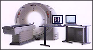
The scanner is connected to a work station that looks similar to a personal computer monitor. With the click of a finger at the computer imaging work station, the technician or doctor can take a closer look at any part of a patient's body. They can rotate the image to see different angles and slice inside an artery for a closer look. The work station is connected to the St. Cloud Hospital's radiology department on a dedicated line. While the patient is still on the table, his image is being transmitted and the doctor in Paynesville and the radiologist in St. Cloud can be looking at the same image instantaneously.
Dr. Bryan Brindley, regional diagnostic radiologist from St. Cloud Hospital, is excited about working with this project as St. Cloud does not have this technology. "I will be able to read the images within five minutes of receiving them and swing my chair around and tell the physician on the telephone what I see," Brindley said.
"The time factor is important in most cases. If we find an abnormality in the patient that needs urgent care, we can airlift the patient to St. Cloud or the University of Minnesota Hospital for faster treatment," Dingmann said.
Ryan Hennen, Vital Images, said medical web browsers can pick up the images anywhere, across the street, city, state, or world.
"It only takes three minutes to create the 3-D image. By rotating the picture, technicians get a more realistic image. Figuraftively, the technician can fly through the aorta vessels from a patient's chest to where it splits at the pelvis," Hennen added. By clicking on the mouse, a technician can change sides from a liver to a heart tissue dissection.
"For a patient comfort level doctors can show their patients the scans, even send a copy home with them," Hennen said. Doctors can e-mail the images to various doctors for consultation, he added.
Following the demonstration, one technician said the scanner will enable the physicians to locate hard to find pathology because of the 3-D images.
The new CT scanner will also enable the hospital to provide a new services for the community. Procedures available include angiography, cardia scoring, and vertiro colonoscopy, according to Dingmann.
The current way of doing an angiography is to insert a catheter into the groin area and injecting a contrasing dye. Patient recovery requires the patient to lay flat on their back five to six hours after the exam.
The CT angiography takes 10 minutes to perform, the contrast injection takes place in your arm, and no recovery time is needed.
CT cornary artery scoring is a study evaluating the cornary arteries in your heart. Patients with calcium in their arteries will show up and they will be scored to see if narrowing of the vessel which can cause heart attacks.
Vertiro colonoscopy is a study by which the CT scanner is used to inspect the colon. In a regular colonoscopy, a scope is used to check the colon and is more invasive. In a CT colonoscopy, the same prep is used, but no scope is inserted in the colon, making it less invasive and less painful .
The procedures will be covered by insurance as long as a clinic requests it for a clinical diagnosis. People who want a cardiac scoring done for personal reasons will be required to pay for the procedure.
Dingmann is excited about the new scanner, giving the hospital new capabilities they have never had before. "The future is now," he added.
PAHCS employees Troy Mansell, CT specialist, and Laurie Meyer, will be spending one week each training in California on the use of the new scanner. The other technicians will receive in-house training.
"Obtaining this scanner has been a nine-month process," Dingmann said. Anyone wishing more information on the scanner can contact Dingmann, 320-243-7711.
The new CT scanner will arrive May 1 and is expected to be operational by May 8.